About
At American Health Imaging, we’ve invested in the most up-to-date imaging equipment to enhance the patient experience and provide the highest quality diagnostic images. These same technologies also allow for greater patient comfort and faster exams, which means your referring physician will get the answers they need… quickly. When you’re dealing with an accident, injury or chronic disease, it’s reassuring to know the efficient workflow at American Health Imaging will have you in / out and results to your doctor in a couple of days, not weeks!

State-of-the-art MRI Equipment
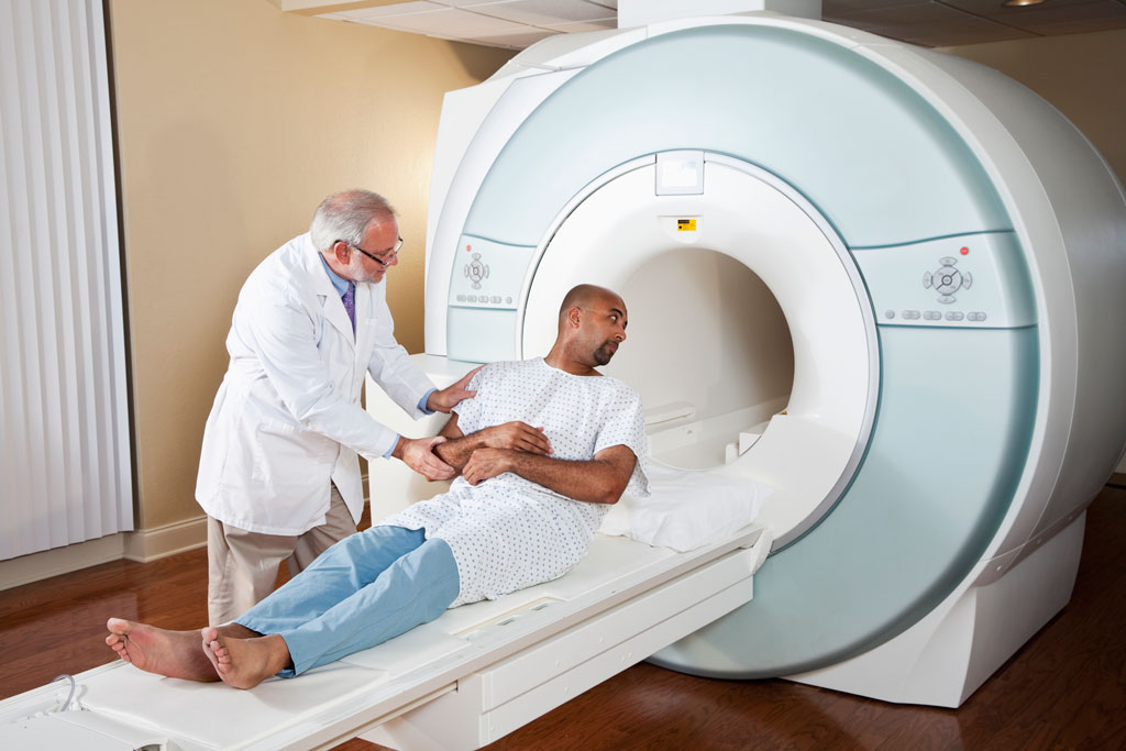
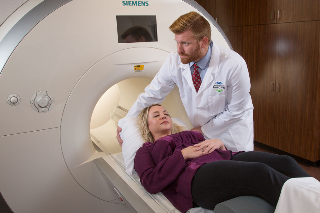
1.5T and 3.0T High-Field, Wide-Bore MRI
These machines boast the strongest magnet field strength available in today’s healthcare environment, offering patients the widest possible range of diagnostic services.

Neurologic Brain Evaluations
The high field strength MRI technology offers crystal-clear visualization and identification of brain tumors, epileptic foci and vascular disease. According to recent studies, the machine also even a 21% increase in the detection of multiple sclerosis lesions compared to standard-strength MRIs.

Orthopedic Extremities Assessments
The high field strength MRI technology allows physicians to view cartilage, tendons, ligaments and bony structures in the fine level of detail necessary to diagnosis injuries and diseases in the arms, hands and fingers. Ultimately, this high resolution enables definitive diagnoses and more effective, lower risk orthopedic surgeries.

Superior Full-Body Exams
The high field strength MRI technology eliminates the need for breath-holding and offers the safety, speed and high throughput necessary for routine abdominal screenings. With its high field strength, the machine can capture the fine details necessary to identify small lesions in the intestines, breast and prostate, including peripheral vascular disease.

Stand-Up MRI
Stand-up MRI allows our patients to be scanned in the specific positions they experience their pain – not just lying down. It also allows all body parts to be imaged with the spine and joints in their natural weight-bearing positions. This open comfort is especially good for people who are super claustrophobic. Patients especially like the distraction of watching TV during their MRI exam at our Stand-up MRI center.

16-Slice Low Dose CT
The dose reduction feature found in the CT at American Health Imaging automates the dose according to patient size, weight, and anatomy, providing high-quality images at minimally required dose.
Our new modern CT system design allows for excellent patient access and comfort. Abdomen scan can be completed with short breath-hold requirements yielding highly detailed images. The fast, minimally invasive vasculature evaluation of the head, neck, thorax, abdomen, pelvis, and extremities using CT Angiography for the quality radiologist are looking for in diagnostics and research.

State-of-the-Art Service
- Physician portals allow you to schedule your exam before you leave your doctor’s appointment
- Leverage our network of hundreds of specialized, board-certified radiologists
- All AHI facilities are ARC- and IAC-accredited, ensuring the highest safety and quality standards

Cutting-Edge Software
- WARP metal artifact reduction is a revolutionary technology that allows patients with metal screws, plates and joint replacements to undergo safe, effective MRI exams.
- BLADE motion reduction technology acts similarly to the image stabilizers on digital cameras. This software accounts for rhythmic motions, such as inhalation, exhalation and heartbeats, to produce more accurate images.
- DTI (Diffusion Tensor Length) is an MRI-based neuroimaging technique that allows radiologists to estimate the location, orientation and anisotropy of the brain’s white matter. This technology makes it possible to diagnose traumatic brain injuries, identify vascular disease and detect brain tumors, multiple sclerosis and other lesions.
- Susceptibility-weighted Imaging level
